
Granulation Tissue After Wisdom Tooth Removal: Normal?
Seeing a pale, bumpy patch after wisdom tooth removal can be unsettling—and it is often normal. Dentists call it granulation tissue. Tiny new blood vessels run through it as the socket starts to rebuild after the clot forms. The shade may drift from pink or red to cream or white. What the color cannot do is diagnose the socket for you.
Start with the trend, not the photograph. Is each day a little easier, or is the pain gathering strength? Fever, drainage that looks like pus, swelling that keeps expanding, and trouble breathing or swallowing are not photo-comparison problems. Use the directions you took home and call the treating office—or our emergency dental team—when the recovery turns the wrong way.
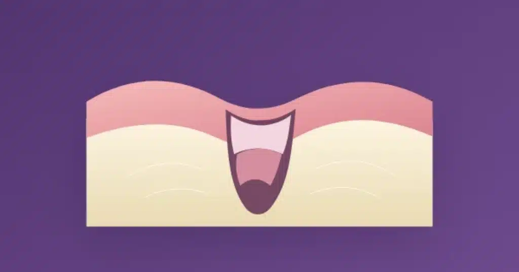
What Granulation Tissue Is Doing in the Socket
Removing a tooth leaves an opening through gum and bone. The clot covers it first. Then cells and fine new blood vessels work their way into the space, building a temporary repair bed—the tissue dentists call granulation tissue.
For a while, it will look nothing like the gum next door. Moist, lumpy, pale: all are possible. The edges draw inward and the outline keeps changing. A tidy surface only tells you about the surface; the bone underneath has much more work left.
This Repair Tissue Is Not Unique to Wisdom Teeth
Front tooth, premolar, molar, wisdom tooth—the repair sequence is much the same. Lower wisdom teeth often leave a deeper hole, so patients notice and worry about those sockets more. The depth makes them awkward to see and clean; it does not create a special kind of tissue.
Root shape, surgical versus simple removal, and the way the gum was managed all change the view. A visible opening may have healthy repair underneath. A closed-looking opening may still have months of bone change ahead.
A Healing Timeline Without False Deadlines
Two sockets rarely keep identical calendars. Upper or lower tooth? Simple removal or surgery? Those differences matter, as do smoking, medication, health conditions, and what happens during aftercare.
Four snapshots—not four deadlines
The first 24-48 hours
The clot is doing the protective work. Leave it undisturbed and use the instructions you were given.
During week one
Repair tissue starts occupying the space. The useful sign is a downward trend in soreness and swelling.
Across the next few weeks
Soft tissue matures and the gum edges creep inward. It may still look different from the surrounding gum.
For months afterward
Below the closed surface, bone formation and remodeling continue quietly.
Use this as orientation, not a deadline for your own mouth. Your treating dentist’s instructions come first.
Is It Healing Tissue, Food, Dry Socket, or Infection?
A picture search is tempting because several things in a socket can look pale, ragged, or dark. Unfortunately, the photo rarely settles it. How the pain and swelling are behaving tells you more, and an examination is the only sensible tie-breaker when you are worried.
White, cream, pale yellow: look beyond the shade
A cream or pale-yellow film may simply be the socket’s protective surface. Thick yellow-green drainage is another matter, particularly beside fever, a strong foul taste, or pain and swelling that are getting worse. Light and saliva distort color easily. The symptom trend is harder to misread.
A dark socket has more than one explanation
Dark does not equal dry socket. Dried blood, clot color, a shadow, and nearby food can all look black or gray from above. Leave it alone. Pain or swelling headed upward, fever, drainage, a foul taste, or bleeding that ignores your pressure instructions is the reason to call—not the shade by itself.
The site seems to be settling
Pink, red, cream, or whitish can all fit. Mild soreness is easing rather than flaring, and you are not testing the tissue with a finger or tool.
What to do: Stay with the aftercare plan and watch the direction of change.
Food may be sitting nearby
Lower sockets, in particular, can catch food around the opening. A hard rinse or improvised tool can damage what is healing.
What to do: Clean it only in the way—and on the schedule—your dentist recommended.
The pain suggests dry socket
Significant pain begins or worsens after the extraction and may travel toward the ear or temple. Bad breath, an unpleasant taste, or an empty-looking site sometimes joins it.
What to do: Call the dentist who performed the extraction.
The recovery has turned toward infection
Instead of settling, the pain or swelling builds. Fever, drainage that resembles pus, spreading redness, or feeling ill may join in. When infection led to the extraction, our guide explains what happens to the abscess after the tooth comes out. The color alone cannot settle the question.
Your next step: Call a dentist urgently. Trouble breathing or swallowing means emergency care.
The Difference Between “Watch It” and “Call”
Things that can fit a normal week
- The surface is uneven rather than smooth
- The tissue shifts among pink, red, cream, and white
- Yesterday hurt a little more than today
- The opening narrows slowly, not overnight
Things worth calling about
- Severe pain, or pain that is gaining strength
- Swelling that grows, fever, or pus-like drainage
- Bad taste or odor that lingers with pain
- Bleeding that will not respond to the pressure instructions
- A change you cannot comfortably dismiss
A Photo Cannot Examine the Socket
If the trend worries you, let Comprehensive Dental Care check it in person. We serve Henderson and the Las Vegas area.
Give the Socket Less to Recover From
Hands and tools stay out. Smoking or vaping pulls against healing and raises dry-socket risk. Choose food that leaves the area alone, brush the rest of your mouth, and do not start rinsing or irrigating ahead of your instructions. Different procedures need different timing; a generic schedule online was not written for your socket.
Five uncomplicated aftercare rules
- Protect the clot. Leave forceful rinsing and spitting for later, according to your dentist’s timing.
- Saltwater, a syringe, or another cleaning method starts when your dental team said it starts.
- Pick food that will not scrape or pack into the opening. Brush everywhere else with a light hand.
- Cold packs and pain medicine follow the directions and your own health restrictions.
- If pain, swelling, bleeding, drainage, or fever reverses course, make the call.
Still preparing for the procedure? Start with our wisdom teeth removal service and the practical factors in our guide to wisdom tooth removal cost.
About the Odd Taste, Smell, or Loose Material
An odd taste or faint odor is not surprising when the area is hard to brush. A foul one that refuses to leave, especially beside rising pain, swelling, fever, or drainage, deserves a call. Do not answer it by digging farther into the socket.
Do not deliberately remove healing tissue. A little surface material may shed as the site changes; losing the protective clot early is different and may leave the socket exposed and increasingly painful. Something came out and the pain is now strong or traveling toward the ear or temple? Call the treating dentist. With no new symptoms, leave the site alone and ask the office whether it needs a look.
When a Lingering Bump Deserves a Look
Usually, the tissue simply matures as the socket closes. Sometimes a bump lingers beside irritation, a retained fragment, or another local issue; it may even show up months later. “Post-extraction granulation tissue” and “extraction granuloma” are labels a dentist may use after looking. They are not invitations to diagnose or cut at it yourself. No progress, repeated bleeding, persistent pain, or growing swelling earns an examination.
Medical Sources Behind This Guide
The biology and timing here draw on peer-reviewed reviews of extraction-socket healing and its cellular stages. For patient-facing guidance, Cleveland Clinic covers both recovery after an extraction and the symptom pattern of dry socket.
Questions People Ask While the Socket Is Healing
When should granulation tissue stop being noticeable?
It commonly appears in the first week and changes over the next two to four weeks as gum covers the socket. The biggest visible differences tend to happen during that first month. Bone repair underneath continues for months, and the exact pace depends on the site, procedure, and your health.
The socket looks white. Is that okay?
Quite possibly. A healing surface can be white, cream, pink, or red. Color becomes more concerning when pain or swelling is climbing, fever appears, the odor turns foul, or drainage resembles pus.
Why isn’t the new tissue smooth?
New blood vessels and connective tissue make an irregular surface while they develop. Do not test the bumps with a finger or tool. Call about severe pain, a rising pain pattern, growing swelling, drainage, fever, or any change that unsettles you.
Is there a safe way to hurry healing along?
No shortcut, unfortunately. The useful work is ordinary: follow your instructions, clean the rest of your mouth, eat and drink comfortably, avoid smoking and vaping, and wait to start rinsing until your directions say so.
Does dry socket look different from granulation tissue?
Sometimes, but pain is the better clue. Dry-socket pain begins or grows after the extraction and may travel toward the ear or temple. Bad breath, bad taste, or an empty-looking opening can join it. Severe or rising pain means call the dentist.
Can healthy repair tissue look pale yellow?
Yes. Cream or pale yellow can belong to the protective surface over a healing site. Thick yellow-green drainage is different, particularly beside worsening pain, swelling, fever, or a strong foul taste, and should be examined.
The socket smells or tastes strange. Is that expected?
A mild, unfamiliar taste or odor may appear while brushing around the site is difficult. A foul one that persists alongside increasing pain, swelling, fever, or drainage warrants a call.
Should I pull white material out of the socket?
No. It may be healing tissue, a protective layer, or food sitting nearby, and scraping cannot tell you which. Use the cleaning method and timing your dentist gave you; ask the office to look if you remain concerned.
Still Comparing Your Socket With Photos?
An in-person look can give you an answer based on the tissue, symptoms, and stage of healing.
This article cannot diagnose a socket. Advice for your extraction and health history should come from the dentist who treated you.